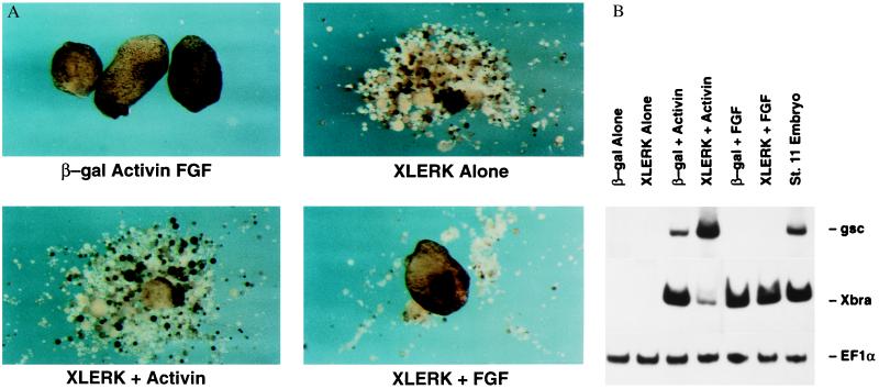
Figure 6.
(A) Morphological analysis of animal caps demonstrating the rescue of XLerk-injected embryos by bFGF. Animal caps were dissected at stage 8.5 and cultured at 23°C in 67% Leibovitz’s L-15 medium, 7 mM Tris⋅HCl (pH 7.5), and 1 mg/ml gentamycin with or without 50 ng/ml Activin or 100 ng/ml bFGF. The animal caps were harvested at stage 26 as determined by uninjected embryos. These results are representative of four independent experiments. (B) Effect of XLerk on gene expression in the animal cap. Embryos were injected with either 5 ng of XLerk mRNA or β-galactosidase mRNA at the 2-cell stage. The animal caps were explanted at stage 8 and cultured in 50 ng/ml activin or 100 ng/ml bFGF until stage 11. Upon harvesting, RT-PCR analysis using primers specific for gsc, Xbra, and EF-1α was performed.

