
FIG 8.
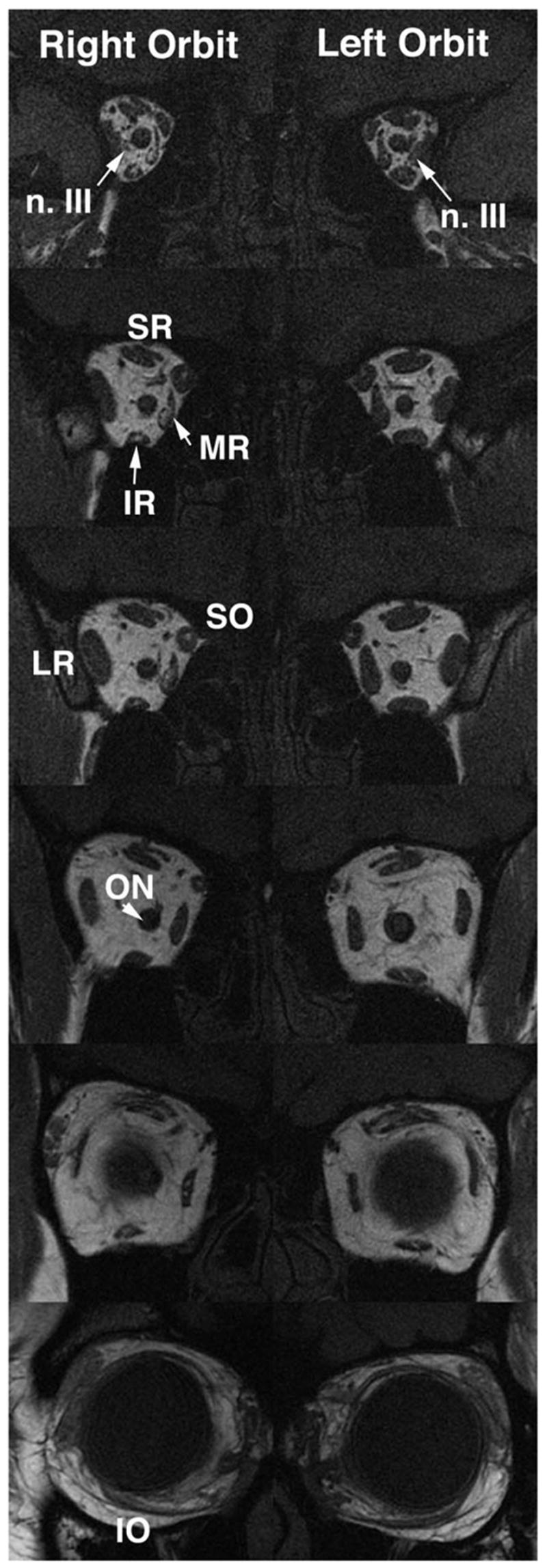
Coronal MRI of both orbits of child with partial congenital right oculomotor palsy. Note hypoplasia and abnormal bright signal in deep portion of the right medial rectus (MR) and inferior rectus (IR) muscles, as well as reduced size of all branches of the oculomotor nerve in the right orbit. Branches of the left oculomotor nerve are also small. Entry of the abducens nerve into the lateral rectus muscle is more posterior to these image planes, and so is not confused with oculomotor nerve branches. The images are 2-mm thick. SR: superior rectus; SO: superior oblique; ON: optic nerve.