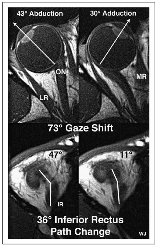
Figure 1. Axial magnetic resonance images (2 mm thickness, T1-weighted) of a right orbit taken at the level of the lens, fovea, and optic nerve (top), and simultaneously in a more inferior image plane along the path of the inferior rectus muscle (bottom), in abduction (left) and adduction (right).

Note the two segments in the inferior rectus path, with an inflection corresponding to the location of the inferior rectus pulley. For this 73° horizontal gaze shift, there was a corresponding 36° shift in inferior rectus muscle path anterior to the inflection at its pulley. IR, inferior rectus muscle; LR, lateral rectus muscle; MR, medial rectus muscle; ON, optic nerve. Reproduced with permission from [13••].
