
Figure 1.
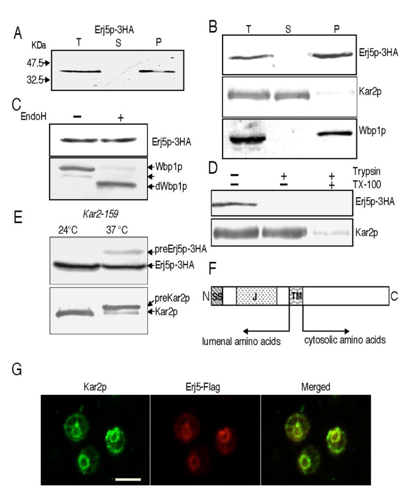
ERJ5 encodes a novel type I ER membrane protein. The Erj5p-3HA protein was detected with anti HA antibodies, the soluble ER lumenal Kar2p and the integral ER membrane Wbp1p were detected with specific antisera. Experiments were performed as described in Materials and Methods. A) Erj5p-3HA is detected in yeast microsomes. Cell homogenates from SSY211 cells that express the Erj5p-3HA fusion protein were centrifuged to obtain microsomal membranes. Supernatant (S) and pellet (P) fractions derived from the equivalent amount of total cell homogenate (T) were resolved by SDS/PAGE and immunobloted for detection of the HA-epitope. B) Erj5p remains in the membrane fraction after treatment of the cell homogenates with 0.1M sodium carbonate pH: 11.5. Supernatant (S) and pellet (P) derived from the equivalent amount of total cell homogenate (T) were immunobloted for detection of Erj5p-3HA. Soluble Kar2p and the integral membrane Wbp1p were immunodetected as controls. C) Erj5p is not N-glycosylated. Microsomes were incubated in presence or absence of Endoglycosidase H before SDS/PAGE and immunodetections of Erj5p-3HA and the Wbp1p glycoprotein as a control. Wbp1p glycoforms containing one or two N-oligosaccharydes or fully deglycosylated (dWbp1p) are indicated with arrows. D) The carboxy-terminus of Erj5p is accessible to proteases in intact microsomes. Three aliquots of yeast microsomes were incubated with or without trypsin in the presence or absence of Triton X-100. Erj5p-3HA and the lumenal soluble Kar2p were immunodetected. E) preErj5p is detected in yeast strains defective in ER protein translocation. A kar2-159 strain expressing the Erj5p-3HA fusion protein (SSY301) was grown to mid-log phase at 24ºC in liquid YPD. Cultures were divided, half was kept at 24ºC, and half was shifted at 37ºC as indicated. After 3 h of incubation, aliquots were removed, cells were lysed with glass beads, and proteins resolved in SDS/PAGE for immunoblotting. Labeled arrows indicate the migration position of preErj5p-3HA and Erj5p-3HA, preKar2p and Kar2p. F) Schematic representation of the overall structure of Erj5p. SS: signal sequence for translocation across the ER; J: J domain, TM: transmembrane region. G) Colocalization of Erj5p and Kar2p in the ER by immunofluorescence. BJ5464 cells expressing Erj5-Flag protein were analyzed by double label immunofluorescence microscopy using anti-Kar2p and anti-Flag antibodies. Left: fluorescent image of Kar2p; middle: fluorescent image of Erj5-Flag; right: merged image. Bar indicates 5 μm.