
Fig. 6.
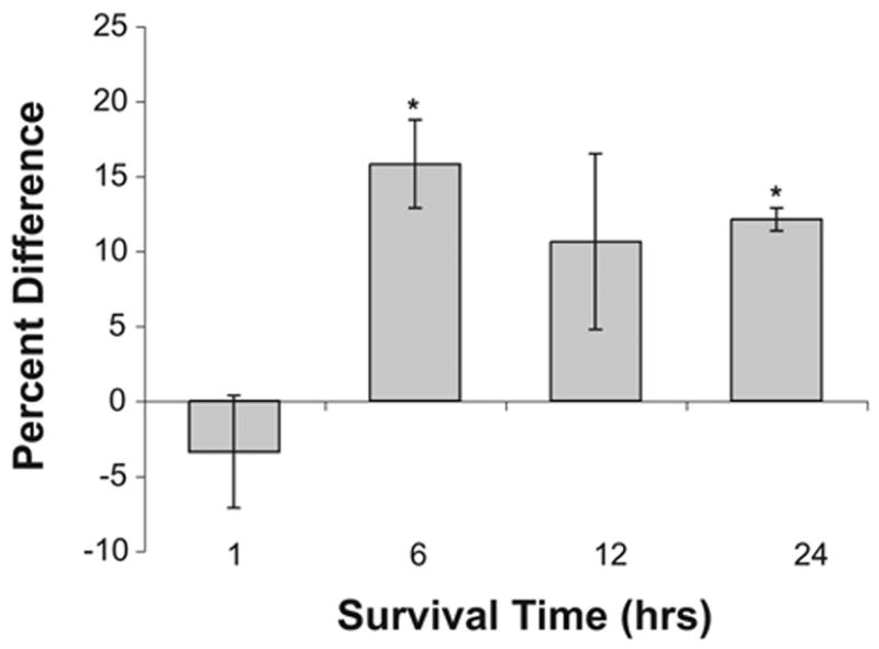
Increased GSTM1 expression following deafferentation. Percent difference in immunolabeling in NM neurons on opposite sides of the same sections measured 1 (n=3), 6 (n=4), 12 (n=3), or 24 h (n=3) following deafferentation. Positive numbers indicate that the deafferented side is darker than the intact side of the brain. Asterisks indicate that there is a statistically reliable difference between the deafferented and intact sides of the brain.