
Fig. 3.
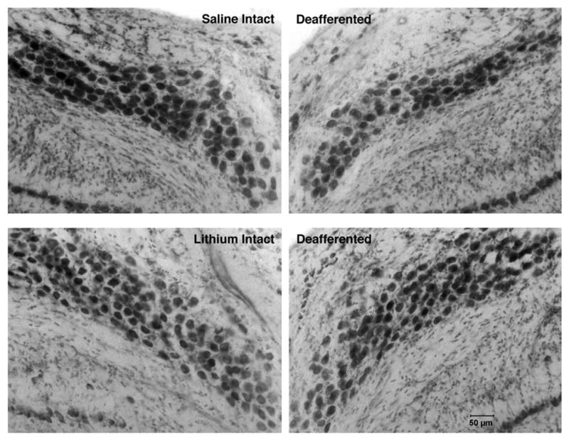
Photomicrographs of intact and deafferented sides of saline- (top) and lithium-treated (bottom) subjects 5 days after cochlea removal. Fewer cells are observed in NM on the deafferented side of the brain. Fewer cells are lost following cochlea removal in the lithium-treated subjects.