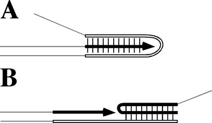
FIG. 8.
Two models of triplex-caused DNA polymerization arrest in vitro. (A) On a single-stranded template, a triplex forms behind the polymerase. The template strand folds back on the newly synthesized strand. (B) On a double-stranded (nicked circular) template, a triplex forms in front of the polymerase. The nontemplate strand folds on the duplex in front when displaced by DNA synthesis. Arrows indicate the direction of DNA synthesis. A triplex is formed within a homopurine/homopyrimidine mirror repeat; the pyrimidine strand is white, and the purine strand is black. Note that a pyrimidine triplex is shown in panel A, whereas a purine triplex is shown in panel B, according to the cited studies (see the text).

