
FIG. 21.
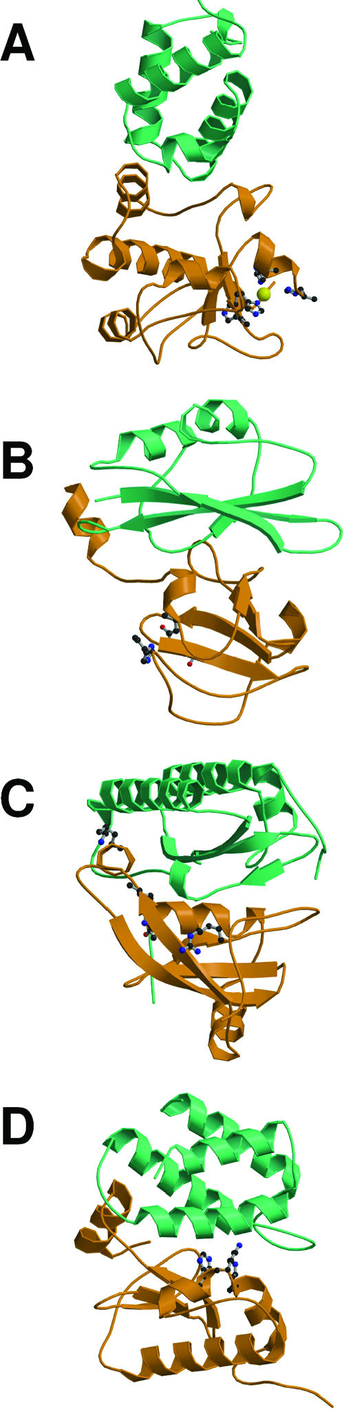
Structures of colicin nuclease domains bound to their cognate immunity proteins highlighting exosite (top) and active-site (bottom) binding by their neutralizing inhibitors. (A) ColE9 DNase-Im9 complex (PDB accession number 1BXI). (B) ColE3 rRNase-Im3 (PDB accession number 1E44). (C) ColE5 tRNase-Im5 (PDB accession number 2FHZ). (D) ColD tRNase-ImD (PDB accession number 1V74). Colicin nucleases are shown in orange, and their Im proteins are shown in cyan. Each structure identifies key active-site residues and, in the case of the DNase, the catalytic metal ion (see the text for details). Courtesy of Irina Grishkovskaya, reproduced with permission.