
Fig. 2. Autoimmune diseases in NF-κB2−/− mice.
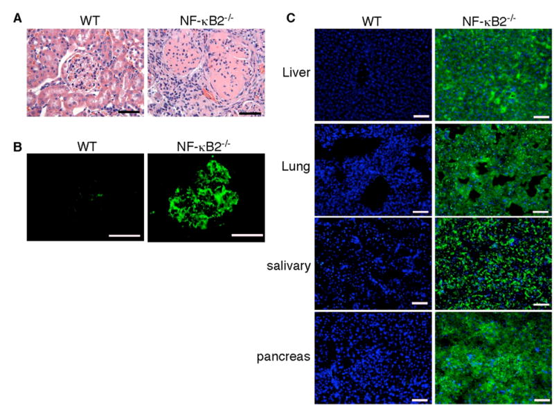
A, H&E staining of formalin-fixed renal sections of deceased NF-κB2−/− and wild-type mice. The renal cortex of the NF-κB2−/− mouse shows mesangial proliferation, crescent formation, and diffuse interstitial lymphocytic infiltrates. B, The presence of IgG-containing immune complexes in glomeruli of the same NF-κB2−/− mouse, revealed by staining cryostat renal sections with FITC-labeled anti-mouse IgG. C, NF-κB2−/− mice have autoantibodies to multiple organs, revealed by FITC-labeled anti-mouse IgM staining of NOD/SCID mouse tissue sections of liver, lung, salivary gland, and pancreas preincubated with serum from individual 1-year-old NF-κB2−/− or wild-type mice. Nuclei were stained with DAPI. Shown is representative staining of the tissue sections with sera from 6 mice for each genotype. Scale bars, 100 μm.