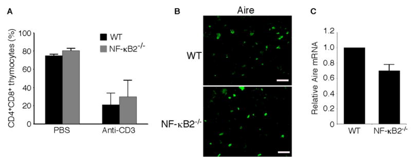
Fig. 3. Effects of NF-κB2 deficiency on activation-induced thymocyte apoptosis and Aire expression.

A, In vivo anti-CD3-induced apoptosis in CD4+CD8+ immature thymocytes in 4- to 6-week-old NF-κB2−/− and wild-type mice. Thymocytes were collected 40 h after intraperitoneal injection of 20 μg of anti-CD3ε (145-2C11) and analyzed by flow cytometry. Data represent means ± standard deviations in percentages of total thymocytes from 3 mice for each genotype. B, Immunofluorescence staining for Aire expression of cryostat thymic sections from 4-week-old NF-κB2−/− and wild-type mice. Shown is representative staining of thymic sections from 3 mice for each genotype. Scale bars, 100 μm. C, Real-time PCR analysis of relative abundance of thymic Aire mRNA in 4- to 6-week-old NF-κB2−/− and wild-type mice. Data represent means ± standard deviations from 4 mice for each genotype.
