
Figure 3.
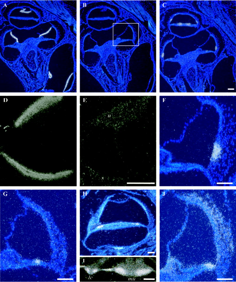
In situ hybridization in cross sections of the mouse inner ear. A) Antisense probe for Vmo1 shows discrete localization of the signal to Reissner’s membrane in cross sections through several cochlear turns. B) Control (sense) probe for Vmo1. C) A similar section to A and B shows hybridization signals obtained using an antisense probe for GM414, a mouse homolog of the zebrafish gene Otolin-1. D) The same probe as in C shows dense signal for GM414 in the maculae of the utricle and saccule. E) Control (sense) probe results for GM414 in the utricle (u) and saccule (s) in a section adjacent to the one depicted in panel D. F) A cross section through one turn of the cochlea at approximately the same level as the boxed region in panel B. Discrete localization of the Scg2 message to cells of the spiral prominence. G) An antisense probe for Umodl1 (olfactorin) reveals discrete localization of the message restricted to the middle of the organ of Corti, possibly in the hair cells or their supporting cells. H) An antisense probe for A430025D11Rik shows hybridization signal in the organ of Corti despite the lack of any signature in the MoCR library. A signature for this gene is found at moderate levels (160 tpm) in the MoVB library. In situ hybridization confirms expression in the vestibular sensory epithelia (data not shown). I) Cib3 in situ hybridization signal in a lateral crista ampularis (lc) and the macula of the utricle (mu). The intense signal on the slopes of the crista and faint signal at the apex is consistent with either supporting cells or hair cells. J) widespread in situ hybridization signal for antisense probe to the novel gene 1110017I16Rik. Scale bars = 100 μm.