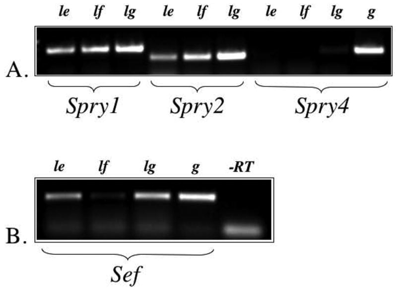
Figure 1.

Expression of Spry1, Spry2, Spry4 and Sef using RT-PCR. A. Spry1 and Spry2, but not Spry4, were detected in the rat lens epithelium (le) and lens fibre cells (lf). Lung tissue (lg), and genomic DNA (g) were used as positive controls. B. Sef was detected in the lens epithelium, lens fibre cells (weakly) and lung. Genomic DNA was used as a positive control, and reverse transcriptase (-RT) was omitted to control for DNA contamination of RNA samples.
