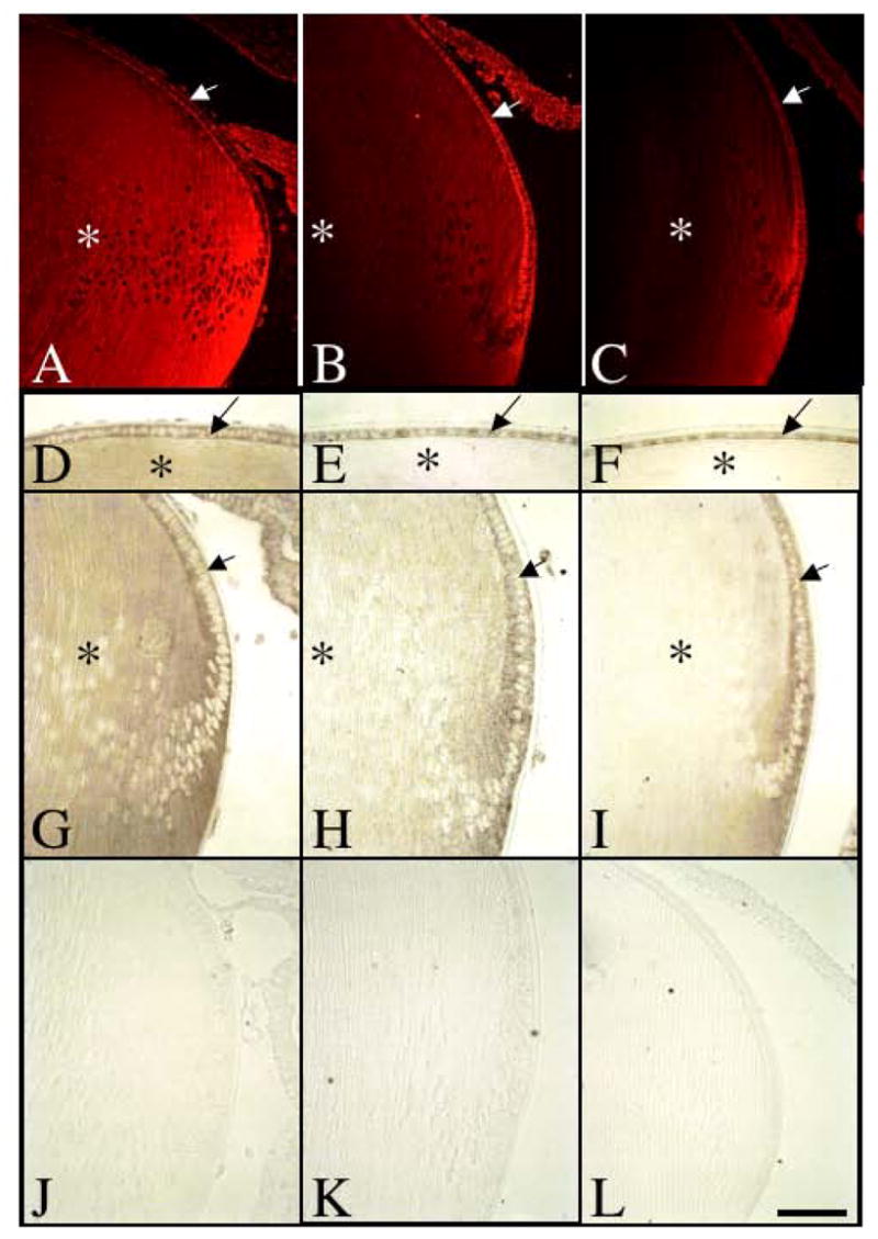
Figure 5.

Expression of Spry2 mRNA during postnatal murine lens growth. Sections of eyes at postnatal stages P3 (A, D, G, J), P15 (B, E, H, K) and P21 (C, F, I, L) were labelled for Spry2 transcripts using either fluorescent in situ hybridisation (A–C) or alkaline-phosphatase labelled riboprobes (D–L). At P3, Spry2 was expressed throughout the lens, in both epithelial cells (A, D, G, arrows) and fibre cells (A, D, G, asterisk). At P15 and P21, Spry2 expression in the lens epithelium (B, C, E, F, H, I, arrow) was retained, but progressively lost in the maturing lens fibre cells (B, C, E, F, H, I, asterisk). Control sections using a sense-labelled riboprobe showed no signal (J–L). Scale bar: 100 μm.
