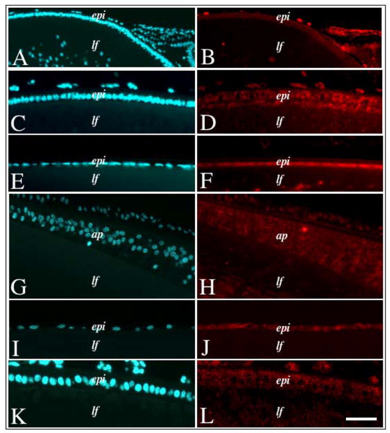
Figure 9.

Immunofluorescent labelling of Sef in lenses of different species. Lens sections from neonatal mouse (A, B), neonatal rat (C, D), weanling rat (E, F), newborn chick (G, H), adult bovine (I, J) and foetal human (K, L) eyes, either immunolabelled for Sef protein (B, D, F, H, J, L) or counterstained with Hoechst dye (A, C, E, G, I, K). In all vertebrates examined, Sef protein labelling was strongest in the epithelial cells with little to no labelling in the fibre cells. Abbreviations: epi, lens epithelium; lf, lens fibres, ap, chick annular pad. Scale bar: 100 μm (A, B), 50 μm (C–L).
