
Figure 2.
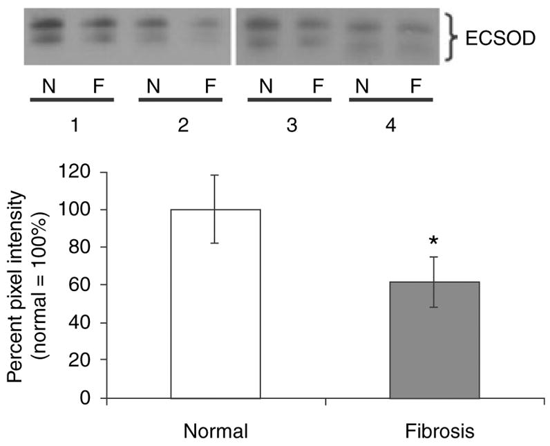
Western Blotting of normal alveolar parenchyma and fibrotic lung parenchyma from usual interstitial pneumonia (UIP) specimens. Homogenates of normal (N) and fibrotic (F) regions from four UIP specimens (nos 1–4) were subjected to reducing SDS–PAGE and Western blotting with an antibody specific for extracellular superoxide dismutase (ECSOD). Both the cut and uncut forms of ECSOD are shown. Densitometry was performed and results expressed as a percentage of normal (normal = 100%). *P = 0.021, Student’s paired t-test, two-tailed.