
Figure 7.
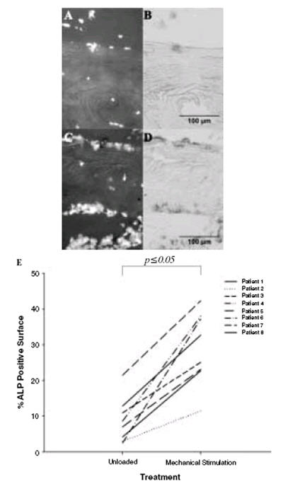
Alkaline phosphatase (ALP) positive bone surfaces. (A) (C) The total cells present are stained using DAPI. (B) ALP was stained using standard techniques and can be seen to be almost absent in a representative image from an unloaded bone sample (D) ALP clearly visible in a loaded sample. (E) Quantification of the percentage ALP positive bone surface revealed an increase (as indicated by the line joining individual patients between treatment groups) in this marker of osteogenic activity in all mechanically stimulated patient cores relative to the unloaded cores.