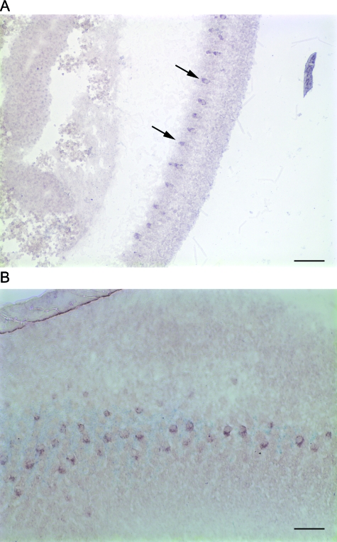
Figure 3.
Iocalization of mRNA encoding AmPNR-like in the pupal head. A. Transverse section of a developing compound eye hybridized with probe for AmPNR-like (arrows); the most intense signal is restricted to a row of large cells along the proximal portion of the retina. The developing optic lobe is visible to the left. Scale bar = 100 m. B. Sagittal section hybridized with the same probe as in A. Scale bar = 50 m.

