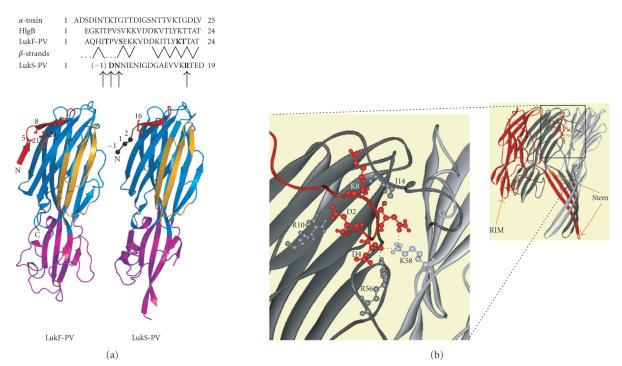
Figure 1.
Structural features of the N-terminal extremities of staphylococcal bipartite leukotoxins and α-toxin. (a) Sequence alignment of the N-termini of staphylococcal α-toxin and of the two components LukF-PV and LukS-PV of the Panton-Valentine leucocidin. The strands arrangement of LukF-PV is indicated by dashes. Cysteine-substituted residues, indicated in bold in the sequences, are also shown on the 3D structures of LukF-PV (PDB code 1pvl) and LukS-PV (PDB code 1t5r). (b) For comparison, view three protomers of the α-toxin heptamer (PDB code 7AHL) and polar interactions involving the N-terminal extremity of a given subunit (red) with residues of two adjacent protomers (grey and dark grey) within the α-toxin pore lumen.

