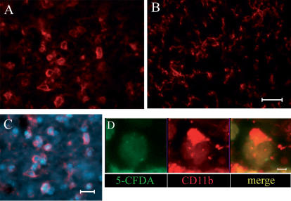
Figure 10. Morphology of microglia/macrophages (red).
CD11b immunopositive cells at day 1 after ischemia and NC(4 h) infusion display a round, phagocytic morphology in the injected (A) but not in the contralateral side were no NC are present (B). Microglia/macrophages appear to surround NC as evidenced in C were NC are marked with Hoechst (blue). A further detail of the interaction between these cells is provided in D where one microglia/macrophage appears to cap a NC (marked with 5-CFDA, green). Bar (A–C): 20 µm; bar (D): 7 µm.

