. 2006 Nov 14;3(1):51–59. doi: 10.1039/b613673g

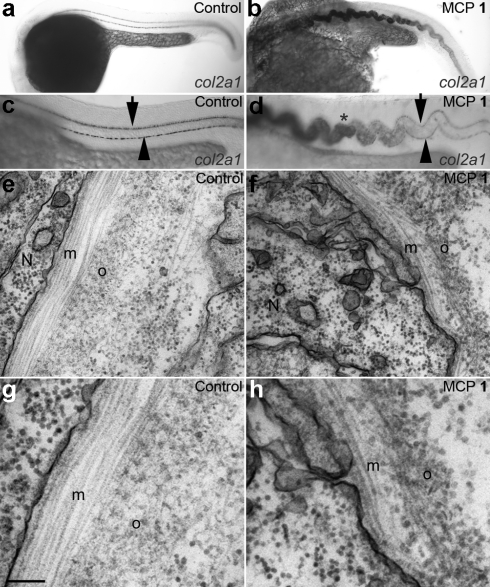
Fig. 6. MCP 1 treatment disrupts notochord sheath differentiation. (a–d) col2α1 is expressed ectopically in the notochord of MCP 1-treated embryos. (a,c) Lateral views of untreated embryo at 24 hpf show lack of expression of col2α1 in notochord throughout trunk and most of tail, contrasting with strong expression in floorplate (arrow) and hypochord (arrowhead). (b,d) Sibling embryo treated with 100 µM MCP 1 shows strong ectopic expression of col2α1 in trunk notochord (*), as well as floorplate and hypochord expression. Identical phenotypes were seen with 2, 3 and 4 (data not shown). (e–h) Transmission electron micrographs of notochord sheath in 27 hpf control (e,g) and MCP 1-treated (100 nM, f,h) embryos. Note regular organization of collagen fibres in medial (m) and outer (o) layers of normal sheath, and disorganized fibre arrangements in MCP 1-treated fish. N: notochord cell. Scale bar, 200 nm (e,f); 100 nm (g,h).

Fig. 6