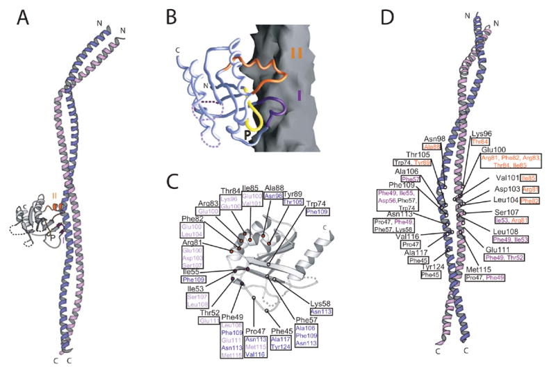
Figure 1.

Structure of the Rab GTPase Sec4p in complex with the GEF domain of Sec2p. (A) A ribbons diagram. Sec4p is grey and Sec2p purple/lilac. Switches I (indigo) and II (orange) and the P-loop of Sec4p are labeled. (B) Sec4p as a Cα worm on the Sec2p surface. (C) Residues of Sec4p within 4 Å of Sec2p are labeled. Residues contacted in Sec2p are boxed and purple or lilac, depending to which Sec2p monomer they belong. (D) Residues of Sec2p within 4 Å of Sec4p are labeled. Contacted residues in Sec4p are boxed; residues in Switch I and II are indigo and orange, respectively.
