
Figure 2.
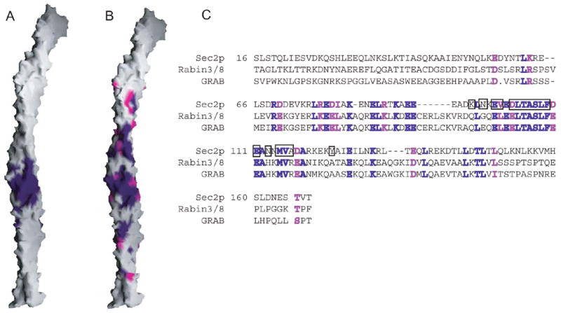
The Sec4p binding surface and conservation. (A) Surface representation of Sec2p. Residues within 4 Å of Sec4p are purple. (B) Residues identical in Sec2p and the mammalian Rabin3/8 and GRAB proteins are purple; similar residues (R=K, D=E, I=L=V) are magenta. (C) Sequence alignment of the Sec2p GEF domain with Rabin3/8 (human isoform β1) and GRAB (Rattus norvegicus). Identical and similar residues are purple and magenta, respectively. The Rabin3 and Rabin8 sequences are almost identical. Residues in Sec2p within 4 Å of Sec4p are boxed.