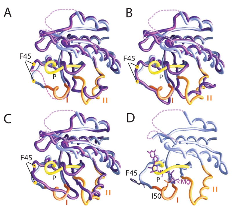
Figure 3.

Superpositions of the Cα backbones of nucleotide/Mg++-free Sec4p from the Sec2p/Sec4p complex (lilac) with nucleotide bound forms of Sec4p (purple) (Stroupe & Brunger, 2000). In the nucleotide-free Sec4p from the complex, switches I and II and the P-loop are labeled, and it is superimposed with: (A) the GDP-bound form. Yellow circles indicate the Cα positions of Phe45. (B) an additional GDP-bound form of Sec4p. (C) the GppNHp-bound form. The conformation of switch II in nucleotide-free Sec4p from the complex more closely resembles that of the GppNHp-bound form than that of the GDP-bound form. (D) Nucleotide-free Sec4p with the positions of GppNHP and Mg++ derived from the GppNHp-bound Sec4p structure indicated. Phe45 and Ile50 are indicated.
