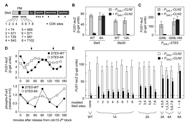
Figure 2.
- Locations of potential CDK phosphorylation sites (SP or TP) in Ste5.
- Elimination of 8 N-terminal CDK sites in Ste5 (Ste5-8A) causes resistance to Cln2.
- Response to α factor was measured in ste5Δ ± PGAL1-CLN2 cells expressing Ste5 variants (WT or 8A) from the native STE5 promoter, or in ste20Δ ± PGAL1-CLN2 cells expressing Ste20 variants (WT or 13A) from the native STE20 promoter. Bars, mean ± SD (n = 8).
- CDK resistance caused by 8A mutations restores membrane signaling independent of Ste5-Gβγ interaction. Gβγ-independent signaling was activated by PGAL1-STE5-Q59L ± 8A in ste4Δ ste5Δ cells ± PGAL1-CLN2. Bars, mean ± SD (n = 7).
- The Ste5-8A mutant disrupts cell cycle periodicity of pheromone response. Cells (cdc15-2 or cdc15-2 STE5-8A) were synchronized in late M phase by arrest at 36°C, and then transferred to 25°C. At various times, response to brief treatment with α factor was monitored (see Experimental Procedures). Top, FUS1-lacZ induction (mean of 4 trials). Bottom, Fus3 activation (phospho-Fus3) was measured using phospho-specific antibodies (mean of 6 trials). Arrows mark the times of bud emergence (see Figures S1D and 5G).
- Ste5 phosphorylation sites were replaced with Ala residues either singly (1A) or in various combinations (2A, 3A, 4A, 8A). Response to α factor was tested in ste5Δ strains ± PGAL1-CLN2 (mean + SD, n = 8-16).

