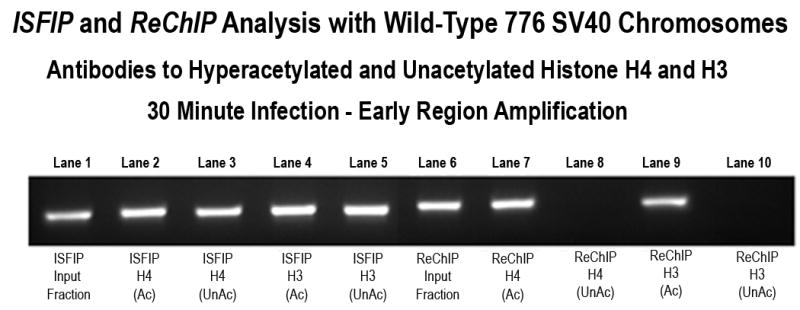
Figure 4A. Presence of unacetylated and hyperacetylated histone H3 and H4 on the SV40 early region in chromosomes undergoing early transcription.

Unfixed SV40 chromosomes were isolated from cells infected with 776 wild type virus for 30 minutes and subjected to an ISFIP/ReChIP analysis with antibodies to unacetylated histone H4 and histone H3 as described in the materials and methods The samples were amplified by simplex PCR with primer sets to the early regions. The position of the amplification product from the wild-type 776 DNA is indicated. Lane 1: ISFIP input fraction; lane 2: ChIP with 7.5 μl of hyperacetylated histone H4 antibody (ISFIP); lane 3: ChIP with 10 μl of unacetylated histone H4 antibody (ISFIP); lane 4: ChIP with 10 μl of hyperacetylated histone H3 antibody (ISFIP); lane 5: ChIP with 10 μl of unacetylated histone H3 antibody (ISFIP); lane 6: ReChIP input fraction; lane 7: ChIP with 7.5 μl of hyperacetylated histone H4 antibody (ReChIP); lane 8: ChIP with 10 μl of unacetylated histone H4 antibody (ReChIP); lane 9: ChIP with 10 μl of hyperacetylated histone H3 antibody (ReChIP); lane 10: ChIP with 10 μl of unacetylated histone H3 antibody (ReChIP). The PCR products in the ISFIP and ReChIP lanes were amplified from one half of the total amount of DNA obtained from each of the samples. The PCR products in the input lanes were amplified from one fourth of the total amount of DNA present in each of the input samples. Similar results were obtained from at least three separate preparations of SV40 chromosomes at 30 minutes post-infection.
