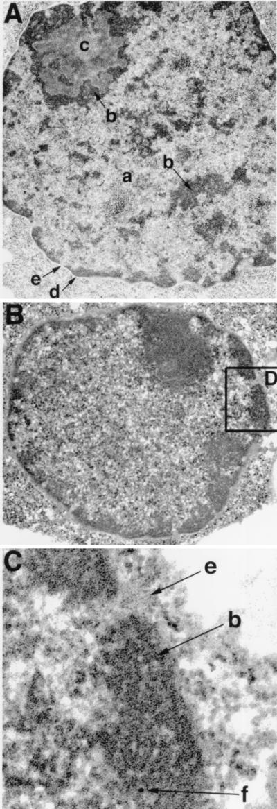
Figure 5.
Ikaros localization in heterochromatin of pre-B lymphocytes. (A) One representative nucleus from the Abelson-transformed pre-B cell line, bcl2–3, is shown at a magnification of 15,000×. The euchromatic (a), heterochromatic (b), and nucleolar (c) regions are indicated along with the nuclear envelope (d) and a nuclear pore (e). (B, C) Low (15,000×) and high power (31,000×) images of a nucleus stained with the anti-N-terminal Ikaros antibody. Localization of one 10-nm gold particle to heterochromatin associated with the nuclear envelope is shown (f).

