Figure 5.

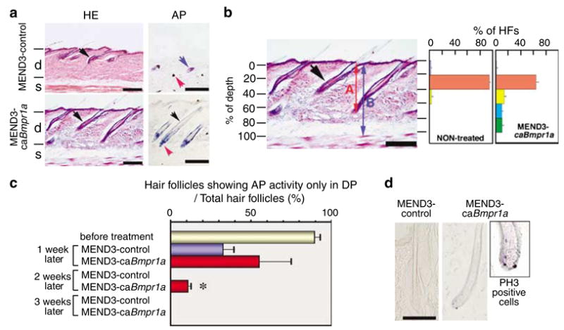
In vivo applications of MEND. (a) Hair follicle formation in mice skin treated with MEND3 containing GFP (MEND3-control) or GFP and a constitutively active form of Bmpr1a (MEND3-caBmpr1a). MEND particles were applied as described in Materials and methods. Tissue sections were stained with HE. Black arrows indicate hair follicles in the dermis. Cells were stained for AP activity to determine hair cycle phase (see text). Blue arrow, sebaceous gland; red arrow, DP; black arrow, ORS; d, dermis; s, subcutis space. Scale bars, 200 μm. (b) A histogram of the number of hair follicles versus depth. Percent depth of hair follicles was calculated by A/B*100 (%). Histograms include averaged data from three controls and three MEND3-caBmpr1a-treated mice. Scale bar, 100 μm. (c) Percent of hair follicles showing AP activity only in DP before and after treatment with MEND3-control or MEND3-caBmpr1a is shown. (d) Cells were stained for PH3 as an indicator of proliferation. Scale bar, 100 μm.
