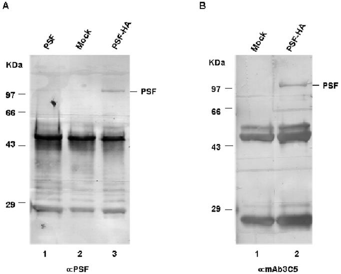
Figure 1.

PSF can be recognized by SR phosphorylation-specific mAb 3C5 in mammalian cells. HA-tagged PSF (pCR3.1·PSF-HA), untagged PSF (pCR3.1·PSF) or the empty expression vector (mock) was transiently transfected into Cos-7 cells. After 48 hrs, supernatants from whole cell lysates were collected and immunoprecipitated with an anti-HA monoclonal antibody. Samples were resolved on 10% SDS-PAGE, transferred to a PVDF membrane, and then western blotting was performed with anti-PSF (A) or mAb 3C5 (B). Bands at ∼50 and 25 kD in all samples result from reaction of the anti-mouse secondary antibody against heavy and light immunoglobulin chains.
