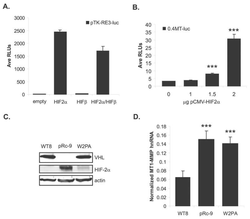
Figure 3.

MT1-MMP transcription is regulated by HIF-2α. (A) WT8 cells co-transfected with pTK-RE3-luc and either pCMV-HIF2α, pCMV-ARNT1/HIFβ, or pCMV-HIF2α + pCMV-ARNT1/HIFβ. Total DNA was kept constant with the addition of pRc/CMV empty vector. Data are of a representative experiment and are presented as average relative luciferase units. (B) Representative experiment of WT8 cells co-transfected with 0.4MT-luc promoter construct and a dose responsive addition of pCMV-HIF2α (mean +/− S.D.). Total DNA was kept constant with the addition of pRc/CMV empty vector; p=0.0001. (C) VHL and HIF-2α protein expression in WT8, pRC-9 and W+2PA cell lines normalized to actin. (D) MT1-MMP hnRNA levels in the three RCC cell lines quantitated by real-time RT-PCR. Values represent the average of pg MT1-MMP normalized to ng GAPDH from ≥3 individual experiments (mean +/− S.D.); p<0.0001. Statistical analyses were performed using the student’s t-test.
