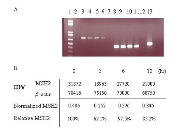
Figure 5.

MSH2 mRNA levels are decreased in MEF WT cells treated with RNAi. A)RT PCR was performed on total RNA extracts from cells co-transfected with pEGFP N3 del1, eGFP3S/47NT, and MSH2 RNAi at different time points after electroporation. Lane 1: 1Kb plus ladder, 2: empty, 3: 0h MSH2, 4: 3h MSH2, 5: 6h MSH2, 6: 10h MSH2, 7: neg control dH20. 8: 0h mus β-actin, 9: 3h mus β-actin, 10: 6h mus β-actin, 11: 10h mus β-actin, 12: neg control dH20, 13: HeLa positive control. B) Spot densitometry was performed to determine the relative percentage of MSH2 present over time after treatment with MSH2 RNAi. Integrated Density Values (IDV) (average density per area) was obtained for both MSH2 and β-actin for each time point and the MSH2 value was normalized to the level of β-actin. The percentage of MSH2 present over time was then determined relative to the zero hour time point.
