
Figure 7.
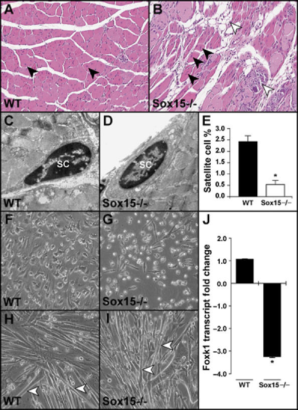
Sox15-deficient skeletal muscle has perturbed regeneration. (A, B) Adult male WT and Sox15−/− mice received intramuscular delivery of cardiotoxin into the tibialis anterior muscles and were killed 2 weeks following injury. Note the centronucleated myofibers (arrowheads) throughout the WT muscle. While the Sox15−/− muscle has evidence of centronucleated myofibers (black arrowheads), there is evidence of impaired regeneration with the presence of fibrous, necrotic tissue (white arrowheads) and adipose tissue replacing myofibers (B; white arrowhead). (C–E) Using electron microscopy, both WT and Sox15−/− tibialis anterior (TA) muscles have evidence of satellite cells (SC), but quantitation (E) reveals a severe decrease in the number of satellite cells in the absence of Sox15. Note the total of nuclei evaluated in WT (1400 nuclei) and Sox15−/− (1096 nuclei) TA muscles (n=3 for each sample; *P<0.005). (F–I) Primary MPCs (including satellite cells) were isolated from postnatal day 2 littermates (offspring from Sox15 heterozygous matings), genotyped and equal numbers of myoblasts were plated. Note decreased numbers of Sox15-null MPCs (G) compared with the WT control (F). Once the respective myoblast cultures were 90% confluent, they were cultured in differentiation media for an additional 3 days. Both the WT and Sox15-null myoblasts differentiated at the same rate and formed equal numbers of myotubes (H and I; white arrowheads) (performed in triplicate). (J) QRT–PCR analysis for Foxk1 expression in WT and Sox15−/− primary MPCs. Note more than a three-fold decrease in Foxk1 expression in the absence of Sox15 (n=3 for each sample; *P<0.005).