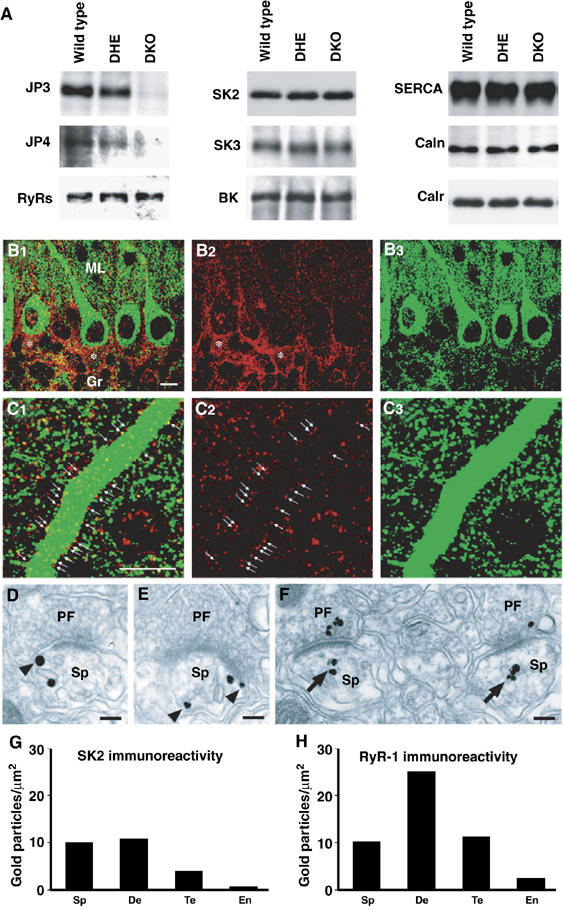
Figure 5.

Expression of functional components for sAHP generation in PCs. (A) Normal contents of major Ca2+ store-related proteins in JP-DKO cerebellum. Total proteins were prepared from the cerebellum and analyzed by Western blotting. Antibodies detected junctophilin type 3 (JP3), junctophilin type 4 (JP4), ryanodine receptor subtypes (RyRs), SK channel type 2 (SK2) and type 3 (SK3), BK/MaxiK channel α subunit (BK), SR/ER Ca2+ ATPase type 2 (SERCA), calnexin (Caln) and calreticulin/calregulin (Calr). In quantitative analysis of immunoreactivities from at least five mice, no differences in protein expression level were observed between the genotypes. (B, C) Double immunofluorescence for SK2 (red) and RyR1 (green). Asterisks indicate SK2 in the pinceau formation, that is, clustered basket cell axons surrounding the initial segment of PC axons. Arrows indicate SK2 on the surface of PC dendritic shafts. Note the presence of dense punctate labeling for SK2 and RyR1 in the neuropil. Scale bars, 10 μm. (D–F) Silver-enhanced pre-embedding immunogold for SK2 (D, E) and RyR1 (F) at synapses between PF terminals (PF) and PC spines (Sp). Note that SK2 is associated with either the cell membrane or the smooth ER (sER, arrowheads), whereas RyR1 is preferentially distributed around the sER (arrows). Scale bars, 0.1 μm. (G, H) Quantitative measurement of the labeling density of SK2 (G) and RyR1 (H) in spines (Sp), dendritic shafts (De), PF synaptic terminals (Te) and capillary endothelial cells (En). Scores on the bars represent the number of immunogold particles per 1 μm2 of given elements. Labeling densities in all of these neuronal elements were much higher than those in capillary endothelial cells indicative of background immunogold decoration. Data were obtained from 8- to 10-week-old mice.
