
Figure 3.
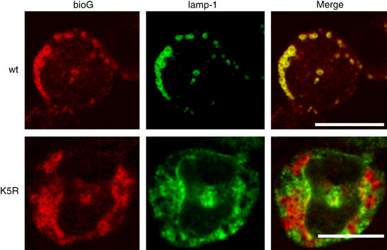
Intracellular localization of K5R G-CSFR in myeloid 32D cells. 32D clones expressing wt or K5R G-CSFR were allowed to bind biotinylated G-CSF (bio-G) for 60 min at 4°C, washed and transferred to 37°C for an additional 60 min. Subsequently, cells were spun on glass slides, fixed, immunostained for bio-G and LAMP-1 and analyzed by CLSM. Scale bar, 10 μm.