
Figure 7.
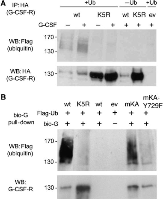
Ubiquitination of G-CSFR. (A) Phoenix E cells cotransfected with HA-tagged wt or K5R G-CSFR and FLAG-tagged Ub were incubated with (+) or without (−) G-CSF for 30 min, and HA immunoprecipitates were analyzed by Western blotting using anti FLAG (upper panel) or anti-HA antibodies (lower panel). (B) A similar setup as in (A), but instead of HA immunoprecipitation, cells were incubated with bio-G for 20 min at 16°C and for 30 min at 37°C and subsequently G-CSFR was pulled down with streptavidin-coated magnetic beads.