
Figure 2.
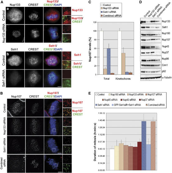
Seh1 RNAi, which prevents the Nup107–160 complex from localizing at kinetochores, or efficient depletion of the Nup107–160 complex, induces a mitotic delay. (A, B) HeLa cells transfected with the indicated siRNA duplexes were pre-extracted before fixation and stained with (Aa) anti-Nup133, (Ab) anti-Seh1 or (B) anti-Nup107 antibodies (red), CREST serum (green) and DAPI (blue). Wide-field microscopy images (A) or maximum projections of deconvolved Z-stacks (B) of representative prometaphase cells and three-fold enlargements of the marked area are shown. Scale bar, 5 μm. (C) Quantification of Nup107 fluorescence intensity in mitotic HeLa cells. Total levels were measured on wide-field images from non-extracted cells (n⩾20) and levels at kinetochores were measured on pre-extracted cells as in Figure 1D. (D) Whole-cell extracts from control, Nup133, Seh1 or ‘combined siRNA'-treated HeLa cells were prepared 3 days after transfection and analyzed by Western blot using the indicated antibodies. α-Tubulin is shown as loading control. (E) HeLa cells treated with the indicated siRNA duplexes were recorded by time-lapse videomicroscopy from 24 to 96 h post siRNA transfection, acquiring images every 5 or 10 min. Duration of mitosis was calculated as the time spent between the first frame where cells rounded up and the first frame where two distinct daughter cells could be observed. Average values of at least 50 cells for each condition are presented. For the complementation experiment (GFP-Seh1siR+Seh1 siRNA), a HeLa cell line stably expressing an siRNA-resistant form of GFP-Seh1 was used (see Supplementary Figure 4B).