
Figure 3.
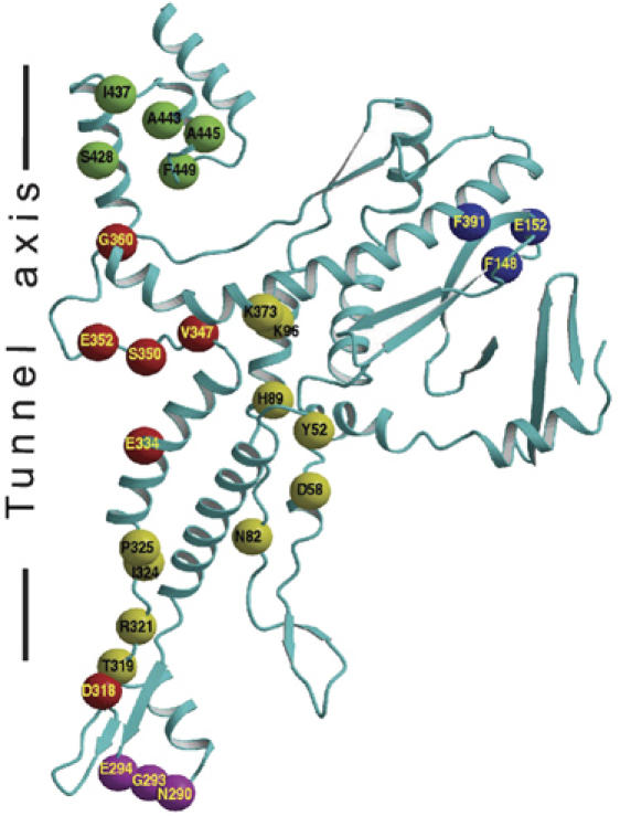
Location of mutations that affected specifically DNA packaging (Isidro et al, 2004a, 2004b). The SPP1 portal protein monomer is shown as a ribbon. The residues, shown as spheres centered at their Cα atoms, are subdivided into five groups: (red) residues in the tunnel loop and residues contributing to the negative charge of the tunnel's surface; (magenta) residues at the base of the portal protein that are likely to interact with the ATPase and gp15; (yellow) residues that could be involved in signal-force transmission between the portal protein and the ATPase and/or carry a structural role; (green) residues involved in stabilization of the crown; and (blue) residues with unassigned function.