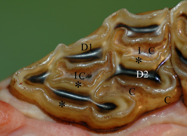
Figure 1.
Normal permanent maxillary P2. Photograph of a P2 occlusal surface. C = cement (light brown), D1 = primary dentin (white/yellowish), D2 = secondary dentin overlying pulp horn (dark brown). * = enamel (visible as a winding ridge). I = infundibulum, (A cone shaped invagination from the occlusal surface of the tooth. The invagination is lined with enamel and filled with cementum (C) to different degrees).8

