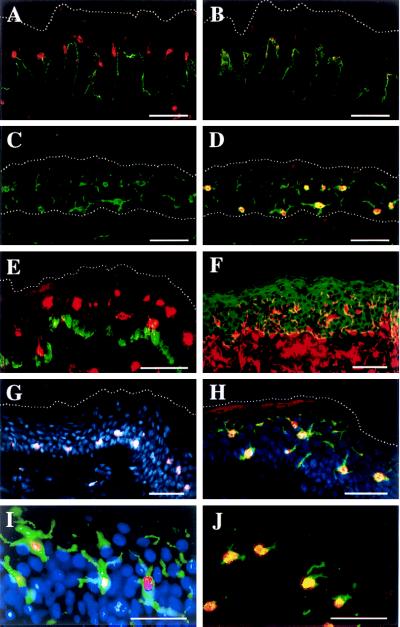
Figure 2.
Immunofluorescence histochemistry localizes 5-LO protein in naïve LCs of normal epidermis. Sample designation: female upper arm, 79 years (C, D, and G–J); male back, 68 years (E); female breast, 29 years (F); female breast, 23 years (A and B). Upper dotted lines (A–E, G, and H) delineate the outer border of stratum corneum. Lower dotted lines (C and D) delineate the border between dermis and epidermis. (A) 5-LO (LO 32 Cy3, red) and collagen VII (Cy2, green). (B) Same as A but antiserum preadsorbed with recombinant 5-LO as described. (C) CD1a Cy2. (D) Same as C plus 5-LO 32, Cy3. (E) 5-LO 1550, Cy3 plus melanin-associated protein, MEL5, Cy2. (F) Vimentin, Texas red plus keratin, fluorescein isothiocyanate green. (G) 5-LO, 1550, Cy3 plus Hoechst 33258, blue. (H) 5-LO LO 32, Cy3 plus CD1a, Cy2 plus Hoechst 33258 blue. (I) Same as H but CCD camera-recorded as described. (J) 5-LO LO 32, Cy3 plus HLA DR, Cy2. (Bars = 50 μm.)

