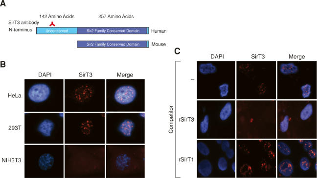
Figure 1.
Full-length SirT3 is nuclear. (A) Schematic representation of full-length human and mouse SirT3. (B) Immunofluorescence analyses of the cell lines indicated using antibody specific to the N terminus of human SirT3. DAP1 was used to visualize the nuclei. (C) HeLa cells analyzed as in B but in the presence of the competitors indicated.

