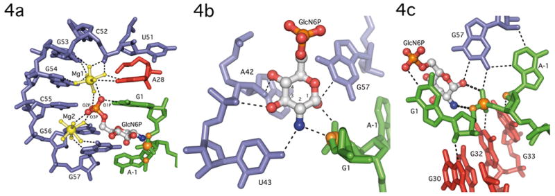
Figure 4.

GlcN6P binding by the GlmS ribozyome. A. Phosphate coordination by two magnesium ions in the GlmS ribozyme. Nucleotides in P2.1 (blue), A28 (red) and G1 (green) use water mediated contacts to organize two hydrated magnesium ions (yellow) and orient the GlcN6P (primarily in gray) phosphate oxygens in the active site. B. Recognition of the GlcN6P sugar ring by nucleobase functional groups. The sugar contacts nucleotides A42, U43 and G57 (blue), and the sugar and 3′-phosphate of G1 (green). The guanine base at G1, which stacks on top of GlcN6P (see Fig. 4a), is not shown to allow all hydrogen bonding contacts to be visualized. The scissile phosphate, 5′-O leaving group and 2′-O nucleophile are shown as orange spheres. C. Active site interactions expected to stabilize this conformation are depicted as thin dashed lines. The catalytically critical interactions between the ethanolamine moiety of GlcN6P and the reactive phosphate are shown as thicker dashed lines. The coloring of individual nucleotides follows the convention in Fig. 3. The scissile phosphate, the 5′-O leaving group and the 2′-O nucleophile (which has been methylated) are shown as orange spheres.
