
Figure 1.
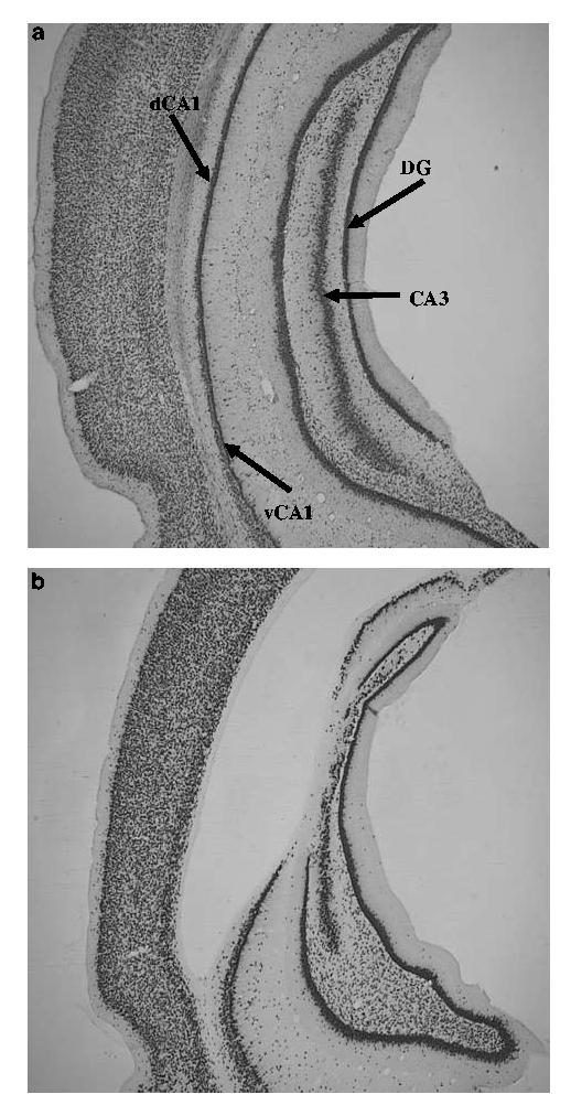
Photomicrographs of representative NeuN-stained sections of adult control (a) and neonatal hippocampal-lesioned (b) rats. Note the neuronal loss in the dorsal CA1, CA3 and dentate gyrus (DG) granule cell layer, and associated ventricular dilation.