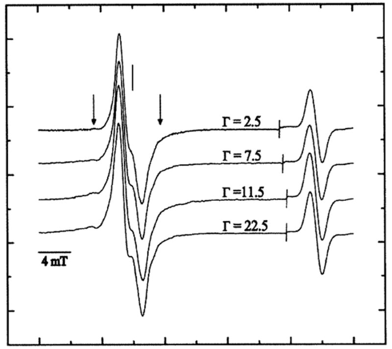
FIG. 2.

First-derivative, Q-band, EPR spectra of pUC18 plasmid X-irradiated with a dose of 90.0 kGy and recorded at 4 K at various Γ. The spectral width was 40 mT. The scan was paused at the line as shown; the field sweep center was then increased by 20 mT and the signal gain adjusted before continuing the sweep to record the strong singlet of the ruby reference at high field. The vertical line corresponds to the position of g = 2.0022. The vertical arrows indicate the broad wing-line features of the putative sugar radicals.
