Figure 1.
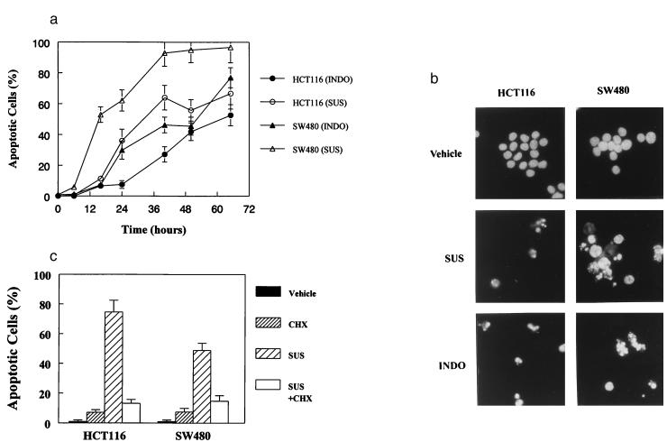
NSAIDs induce apoptosis in human colorectal cancer cells. (A) HCT116 and SW480 cells were treated with INDO (300 μM) or SUS (125 μM for HCT116 and 200 μM for SW480 cells). Cells were collected at the indicated times and scored for morphological evidence of apoptosis as described in Materials and Methods. (B) HCT116 and SW480 cells were treated with SUS or INDO for 60 hr, harvested, and assessed for morphological signs of apoptosis. Cells treated with SUS or INDO show the hallmarks of apoptosis. Independent evidence of apoptosis was obtained by examining phosphatidylserine exposure. SUS treatment of HCT116 and SW480 cells resulted in substantial phosphatidylserine exposure indicative of membrane unpacking and apoptosis. Forty-eight hours after treatment with 125 μM SUS, 44% of HCT116 were apoptotic as judged by phosphatidylserine exposure vs. 4% in the vehicle-treated control cells. Likewise, 74% of SW480 cells were apoptotic 50 hr after treatment with 200 μM SUS vs. 1.5% of the vehicle-treated control cells. (C) CHX inhibits SUS-induced apoptosis in HCT116 and SW480 cells. Cells were treated with SUS as above in the presence or absence of CHX (10 μM). Apoptotic cells were evaluated as described above.

