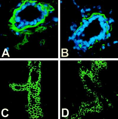
Figure 6.
Immunolocalization of E-cadherin and laminin. Laminin immunoreactivity (A and B), green color (nuclei are blue), in mammary-duct cross-sections circumscribes the mammary epithelium of PR-A transgene-negative mice (A) but is discontinuous and decreased in the gland of PR-A transgenic mice (B). Cadherin immunoreactivity (green color) delineates the epithelial cells in this branching duct of transgene negative mice (C), which is greatly diminished and disorganized in the epithelium of PR-A transgenic mice (D). In all cases, without primary antibody, there was no immunoreactivity (data not shown).

