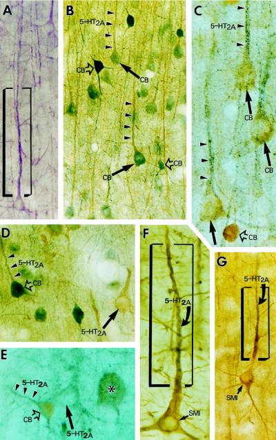
Figure 4.
Color light micrographs demonstrate the accumulation of 5-HT2A receptor immunoreactivity in the proximal dendrites of pyramidal and nonpyramidal neurons in prefrontal area 46. The neurons were neurochemically identified by using 5-HT2A/CB (B–E) or 5-HT2A/SMI-32 (F–G) double immunocytochemistry. Arrowheads point to dendrites, arrows point to pyramidal cell somata, and open arrows point to nonpyramidal cell bodies. (A) 5-HT2A-positive pyramidal cell in layer III (stained purple with the Vector VIP staining kit). (B and C) Two 5-HT2A/CB double-stained sections from cortical layer III demonstrate that 5-HT2A immunoreactivity (arrowheads) is colocalized with CB immunolabel (arrows) in pyramidal neurons. (B) 5-HT2A label is light brown and CB label is bluish gray. (C) Double labeling is “reverse,” 5-HT2A label is bluish gray, and CB label is light brown. (D and E) Two micrographs from layer V demonstrate that large- (asterisk) and medium-size interneurons (open arrows) colocalize 5-HT2A receptor and CB and show that 5-HT2A receptor is also present in CB-negative pyramidal cells (arrows), typical of the infragranular layers. (D) 5-HT2A, light brown; CB, bluish gray. (E) 5-HT2A, bluish gray; CB, light brown. (F and G) 5-HT2A/SMI-32 double-labeled pyramidal cells from layer III. Note that the gray color of 5-HT2A label dominates only in the proximal apical dendrites (within the frame), whereas thinner dendritic branches and the cell body are overshadowed by the brown-colored SMI-32 staining.

