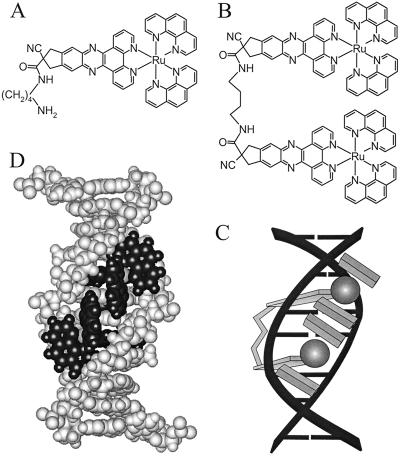
Figure 1.
Structures of 1 (A) and 2 (B) and schematic picture of the binding of 2 (Δ,Δ isomer) to DNA (C). Molecular model of 2 (Δ,Δ isomer) bound to a duplex decanucleotide (D), with the ruthenium centers situated in the minor groove (obtained by energy minimization in the hyperchem software package with the Amber force field).

