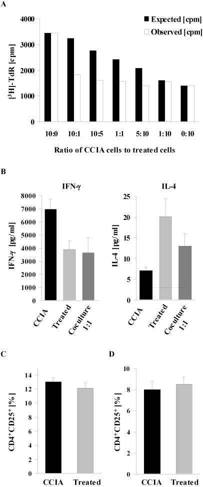
Figure 6. Lymphocytes from epicutaneously immunized mice inhibit proliferation and IFN-γ production by lymphocytes from CCIA mice.
Splenocytes from a sham-treated CCIA control were cocultured with splenocytes from an epicutaneously treated mouse at varying ratios (A). Total number of cells in the culture was kept constant. Cocultures were stimulated with 50 µg CII/ml and CII-specific splenocyte proliferation was assayed as in Figure 3. Black bars demonstrate the expected proliferation [cpm] in the culture, calculated by the formula: [cpm] = a f+b(1−f), where a = [cpm] with 100% control cells, b = [cpm] with 100% treated cells, f = fraction of control cells in the culture. The observed [3H]-thymidine incorporation is shown in white bars and represents the mean of triplicate cultures. Similar results were obtained in four individual experiments. Production of IFN-γ and IL-4 in 1∶1 ratio cocultures of splenocytes from control and treated mice were assessed by ELISA and results are expressed as mean pg/ml+1 SEM (n = 4) (B). No cytokines were produced in response to a control antigen or when cells were not stimulated. The proportion of CD4+CD25+ T cells in freshly isolated splenocytes (C) from CCIA control or epicutaneously immunized mice and after 90 h in vitro stimulation with 50 µg CII/ml (D) were assessed by flow cytometry. Results are expressed as the mean [%] CD4+CD25+ T cells out of the total CD4+ population+1 SEM (n = 9). (▪) = treated with epicutaneous CII, (▪) = sham-treated CCIA controls.

