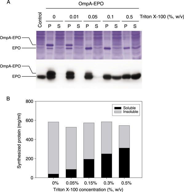
Figure 5.
In situ cleavage of the signal peptides during cell-free protein synthesis. (A) The indicated concentration of Triton X-100 was added to the reaction mixture from the start of the synthesis reaction, and examined to determine it could increase the level of signal peptide cleavage. After incubation, the reaction mixture was centrifuged at 10 000 g for 10 min, and the soluble and pellet fractions were analyzed by 13% Tricine-SDS–PAGE stained with Coomassie Brilliant Blue (upper panel) and western blot using the anti-EPO antibody (lower panel). P and S represent the insoluble and soluble fractions of the cell-free synthesized EPO, respectively. (B) [14C]leucine-labeled radioactivity of the expressed protein was measured as described under Materials and Methods.

