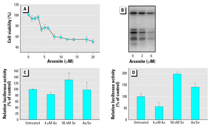
Figure 8.
Arsenite reduces but does not eliminate readthrough of UGA codon in serum-containing medium. To analyze the cell’s ability to readthrough the UGA codon with the appropriate SECIS element, we used a gene reporter fusion described by Handy et al. (2005). (A) Cytotoxicity of arsenite in COS7 cells. MTT dye reduction was assessed after incubating cells with arsenicals for 24 hr, and the absorbance obtained was compared with untreated cells to determine the relative percent cell viability. Error bars represent mean ± SD for at least three independent cultures treated with arsenite. (B) Arsenite blocks incorporation of radiolabeled selenium in COS7 cells. Cells were cultivated in the presence of 75Se-selenite (10 nM) and treated with sodium arsenite at the concentrations indicated. Cells were harvested after 24-hr incubation, and 20 μg of protein from crude cell extract was separated by 12% SDS-PAGE. (C) Readthrough of UGA codon is resistant to arsenite. The gene reporter fusion denoted UGA SECIS/S (Handy et al. 2005) was transfected (250 ng) into COS7 cells cultured in DMEM with 10% serum. Transfection efficiency was determined by co-transfecting pRL-CMV encoding Renilla luciferase (Promega). Relative luciferase activity in control cells (no arsenicals) is taken as 100% readthrough. Selenium and/or arsenite was added to the culture medium before transfection at the concentrations indicated. (D) Readthrough of UGA codon determined in low-serum conditions (0.5% serum). Error bars represent mean ± SD for at least three independent transfected cultures.

