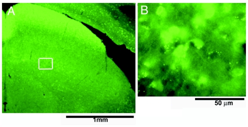
Fig. 1.
GFP-positive neurous in SGI. (A) Low-magnification photomicrograph of the superior colliculus in a GAD67-GFP mouse. (B) High-magnification photomicrograph of neurons in SGI (from A Inset) that express GFP fluorescence. This GFP fluorescence allows selective, visually guided sampling of GABAergic neurons that, because of their small size and number, otherwise are rarely encountered.

